CFA occlusion: an unexpected strategy
A 56-year-old man with multiple cardiovascular risk factors presents with recent-onset rest pain, raising concern for acute limb ischemia on right side.
Imaging reveals extensive multilevel disease, including occlusion at the right common femoral artery and diffuse involvement of the right femoropopliteal and infragenicular segments.
Faced with this complex anatomy and high-risk profile, the key question is clear: which revascularisation strategy offers the best chance to restore flow and preserve the limb?
Several options can be considered, each with its own implications.
What would be your approach?

Patient profile and risk factors
- Bilateral peripheral neuropathy
- Rest pain for the last 24h – Fontaine IIa
- A 56-year-old male patient
- Obese, hypertension, type 2 diabetes (DM 2)
- Bilateral retinopathy,
- Non-dialysis-dependent renal dysfunction,
- Previous myocardial infarction
- 💊 Poor medical adherence
CTA findings
- Common femoral artery (CFA) with signs of occlusion
- Superficial femoral artery (SFA) and deep femoral artery (DFA) with atherosclerotic disease. Occlusion and stenosis of the proximal third of the SFA, with signs of recanalisation in the middle and distal thirds.
- DFA with occlusion and recanalisation in the distal third.
- Popliteal artery with atherosclerotic changes and occlusion.
- Infragenicular arteries show normal anatomical distribution. The anterior tibial (AT), posterior tibial (PT), and fibular arteries show sequential stenoses and occlusions.
Treatment strategy
We decided that the optimal strategy in this case was an initial debulking with atherectomy, followed by a stentless bifurcation-based approach using POBA and DCB angioplasty, with no role for thrombectomy, IVL, or routine stenting.
Key evidence supporting the strategy
Acute arterial occlusion (archived)
StatPearls [Internet]. Treasure Island (FL): StatPearls Publishing; 2026 Jan – . PMID: 28722881.
Smith DA, Lilie CJ.
Cases of limb-threatening ischemia require an emergent vascular surgery consult. The surgical approach focuses on reperfusion of the affected extremity. This can be accomplished by surgical bypass, endarterectomy, or embolectomy. Results are variable and will ultimately depend on the duration of the ischemia and the extent of occlusion. Catheter-directed thrombolysis, performed by an interventional radiologist, is becoming increasingly common and is reserved for patients with a salvageable limb (Rutherford classes II, IIa, and IIb). Patients presenting with profound paralysis and absent pain with inaudible arterial and venous pulses are considered to have irreversible damage and will require amputation. Surgical treatment options are typically reserved for more severe cases or when nonsurgical management is unsuccessful. These options include the following: open bypass surgery; endovascular therapy, such as stenting, balloon angioplasty, or atherectomy.
Importance of successful revascularisation in acute limb ischemia: sub-analysis from the RESCUE ALI trial
Catheter Cardiovasc Interv. 2025 Jan.
Haraguchi T, Tan M, Uchida D, Dannoura Y, Shibata T, Iwata S, Azuma N.
Technical success in ALI treatment significantly enhances 1-year AFS rates. Thus, choosing the appropriate revascularisation procedure based on predictors of technical success is crucial for improving patient outcomes.
Acute ischemia of the upper and lower limbs: tailoring the treatment to the underlying etiology
Semin Vasc Surg. 2023 Jun.
Ferrer C, Cannizzaro GA, Borlizzi A, Caruso C, Giudice R.
Despite the development of several therapeutic options for upper and lower limb acute ischemia, the optimal management remains to be determined. Prognoses for limbs and patients' survival vary according to the accuracy of the evaluation process and the promptness of the therapeutic interventions, including revascularisation and limb amputation. An adequate preoperative assessment, including medical history, time of symptom occurrence, severity of clinical presentation, and etiology of ALI are essential to guide treatment decisions.
Initial approach
- Intravenous fluid resuscitation
- Intravenous analgesia for pain control
- Intravenous anticoagulation with heparin to minimise thrombus propagation
(Therapeutic dosing: low molecular weight heparin or unfractionated heparin)
The general goal of anticoagulation is to increase the activated partial thromboplastin time (aPTT) to 2.0–2.5 times the baseline value.
Percutaneous approach
- Left common femoral artery (CFA) puncture with 6F short sheath.
- Antegrade profunda femoris artery (DFA) recanalisation using 0.018" guidewire and support catheter.
- Exchange for 7F × 45 cm long sheath.
- Antegrade superficial femoral artery (SFA) recanalisation with a second 0.018" guidewire (kissing wire technique) and support catheter.
- Intravascular ultrasound (IVUS): intraluminal positioning, showing minor dissection at the proximal SFA and luminal reduction at the popliteal artery.
- Pre-dilatation with non-compliant balloon (NCB) Jade 4 × 80 mm in CFA / DFA / SFA.
- Atherectomy with JetStream device in CFA / SFA.
- Drug-coated balloon (DCB) angioplasty with Ranger 7 × 120 mm in CFA / SFA. (No frame capture available due to technical issue.)
- Popliteal segment: intra-arterial vasodilator infusion followed by DCB Elutax 4 × 80 mm.
- Final angiography: satisfactory vessel remodeling with good distal runoff.
- Closure of left CFA with Perclose ProStyle device.
Immediate post-procedural follow-up
- 💊 COMPASS regimen: Rivaroxaban 2.5 mg twice daily
- Aspirin 100 mg once daily
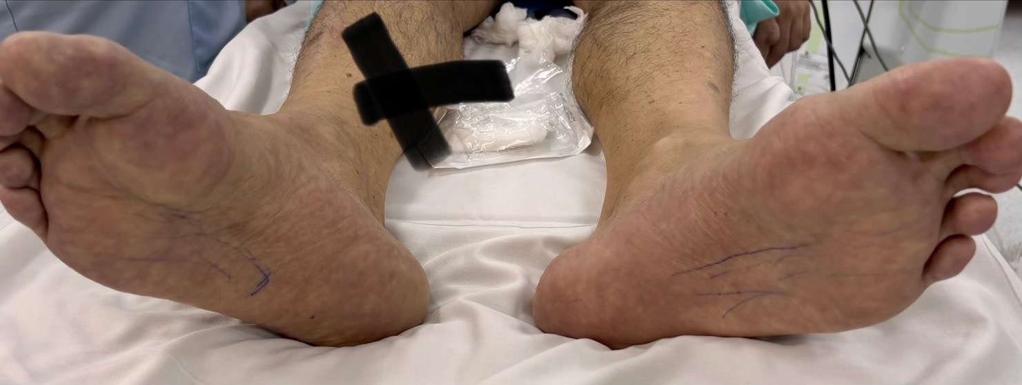
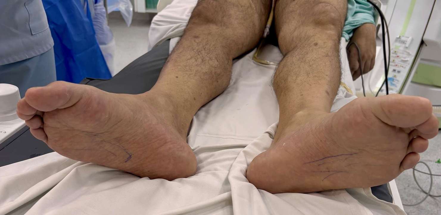
- Palpable pedal pulses
- Warm foot
- (Coronary stenting planned 14 days later)
Duplex ultrasound (Feb 2026)
Triphasic flow patterns with preserved velocities are observed in all evaluated arterial segments, except for the posterior tibial artery, which shows no detectable flow.
Absence of compressibility and absence of flow are consistent with arterial occlusion.
Get the latest clinical cases and breaking news delivered straight to your inbox!
