When a juxtarenal aneurysm raises an infectious concern
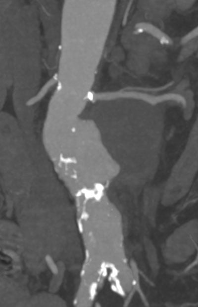
A rapidly expanding saccular juxtarenal aneurysm in an elderly patient, accompanied by unexplained weight loss and an inflammatory response, raises important questions about both its underlying cause and the optimal treatment strategy.
In this clinical case, the multidisciplinary team faced the challenge of managing a 75-mm juxtarenal aneurysm with features raising suspicion of an infectious process.
What treatment strategy would you choose?

A rapidly expanding aneurysm in a symptomatic patient
- A 73-year-old woman with hypertension, type 2 diabetes and chronic kidney disease (eGFR 46 mL/min/1.73 m²) presented with a 7-kg weight loss over 2 months.
- At the same time, her saccular juxtarenal aneurysm had expanded by 2 cm, reaching 75 mm in diameter.
Multidisciplinary evaluation
- The patient was afebrile at presentation.
- Laboratory blood testing showed an elevated inflammatory response, with:
- CRP: 63 mg/L
- WBC: 12 × 10⁹/L
- An extensive infectious work-up was performed, including:
- Four sets of blood cultures
- Pneumococcal urinary antigen testing
- Serologies for Bartonella, Brucella, Q fever and Rickettsia
- Immunological serology
- Chest imaging revealed left lower-lobe pneumonia.
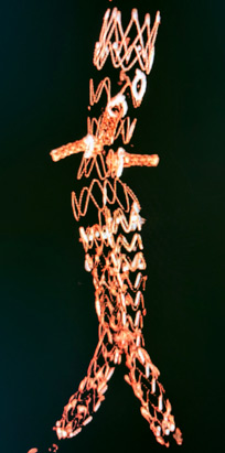
With the clinical and anatomical challenges in mind, the team opted for an off-the-shelf alternative to a custom-made device.
A Terumo aortic TREO device was modified on the back table to create a physician-modified endograft (PMEG) tailored to the patient’s anatomy.
Uneventful postoperative course
The postoperative course was uneventful, with no spinal cord ischaemia, stable creatinine levels and no access-site complications.
The patient spent her first night in the ICU before being transferred to the infectious disease unit on the first postoperative day.
Further management
Following the endovascular procedure, direct sac puncture and embolisation of the aneurysm sac were performed.
Cone-beam CT was used to assess the result, with pre- and post-embolisation imaging demonstrating the changes in the aneurysm sac.
A sac biopsy was also obtained, with the results still pending at the time of reporting.
Get the latest clinical cases and breaking news delivered straight to your inbox!
