When previous FEVAR complicates the picture
A 72-year-old man with a history of fenestrated endovascular aortic repair (FEVAR) presents with abdominal symptoms and multiple comorbidities.
What initially appears to be a non-specific clinical picture soon evolves into a complex vascular challenge, where previous aortic repair plays a pivotal role in determining the treatment strategy.
Faced with several possible therapeutic options, what would you choose?

Clinical presentation
A 72-year-old man with significant cardiovascular comorbidities presented with non-specific abdominal symptoms in the context of previous complex aortic repair.
Medical history:
- Systemic hypertension
- Chronic ischaemic cardiomyopathy
- Atrial fibrillation
- Mild renal insufficiency
- Myelodysplasia
Previous aortic intervention:
- FEVAR performed in 2023
- including four bridging stents: for SMA + CT + 1 RRA + 2 LRA (scallop for CT)
Recent clinical presentation:
Assessment 20 days earlier for abdominal constipation and asthenia
Therapeutic strategy
The discovery of a spontaneous pseudoaneurysm raised the suspicion of an infectious process, requiring further investigation and careful monitoring.
Microbiological assessment:
- Blood cultures and urine cultures performed
- No infectious agent identified
Management approach:
- Prophylactic antibiotic therapy started
- Continuous surveillance of vital parameters and clinical status
Intervention programmation
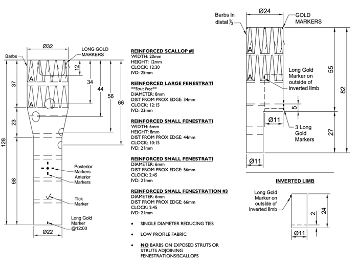
Many technical options have been discussed:
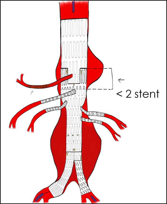
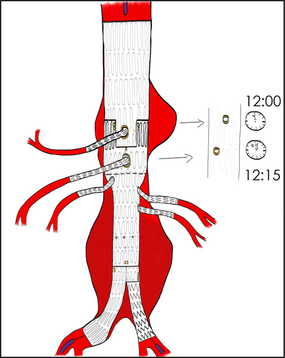
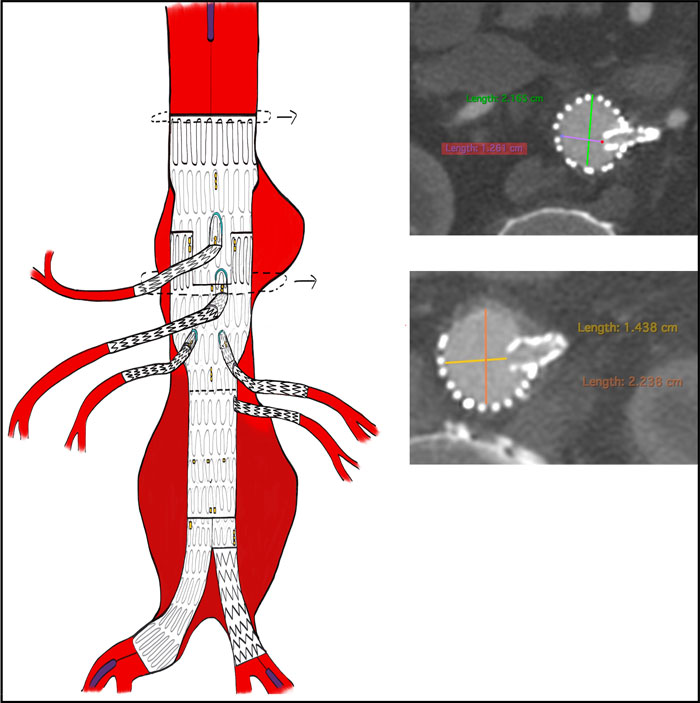
Clinical deterioration
48 hours later, the patient's condition suddenly worsened:
- Acute onset of abdominal pain
- Severe hypotension (70/40 mmHg)
- Acute anaemia
Diagnosis:
Rupture of the pseudoaneurysm leading to haemodynamic instability
Programmation during emergent setting
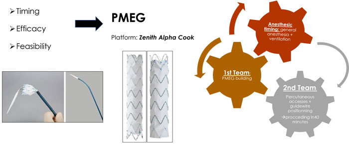
Procedure
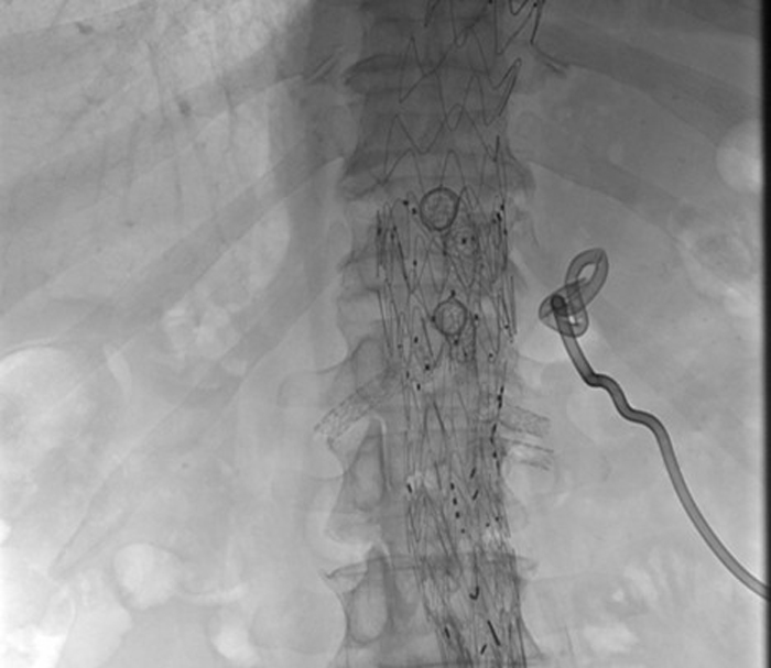
PMEG with 2 fenestrations for CT + SMA to extend above and exclude the aneurysm with sufficient distal sealing in the previous fEVAR.
Following the emergency procedure, the patient was closely monitored in the intensive care unit.
Post-operative care:
- 24-hour admission to the ICU
- Close clinical surveillance
Biological assessment:
- Transient increase in serum creatinine to 2.3 mg/dL
- No fever reported
- Two blood cultures remained negative
Further investigation:
PET-CT scan scheduled to further assess the underlying aetiology
Late post-operative course
After an initially stable recovery, the patient presented 27 days later with new clinical signs raising concern for a potential underlying complication:
- Lumbar pain
- Fever
- Asthenia
Microbiological assessment and ongoing management
Microbiological investigations:
- Blood cultures remained negative
- Collection drainage was sterile
Therapeutic management:
Antibiotic therapy initiated with dalbavancin
Further clinical evolution to be continued...
Get the latest clinical cases and breaking news delivered straight to your inbox!
