Cases and resources in vascular techniques
This section provides a selection of cases and resources provided by experts in vascular techniques.
In conversation with Dr Gustavo Basso, Prof. Sarah Thomis and Prof. Jorinde van Laanen review the latest evidence on compression therapy after endovenous treatment, pelvic venous disorders and medical treatment following endovenous interventions.
Why was a Delphi consensus needed for femoropopliteal artery disease? In this one-minute interview, Prof. Secemsky explains how expert guidance can help navigate the growing range of endovascular treatment options.

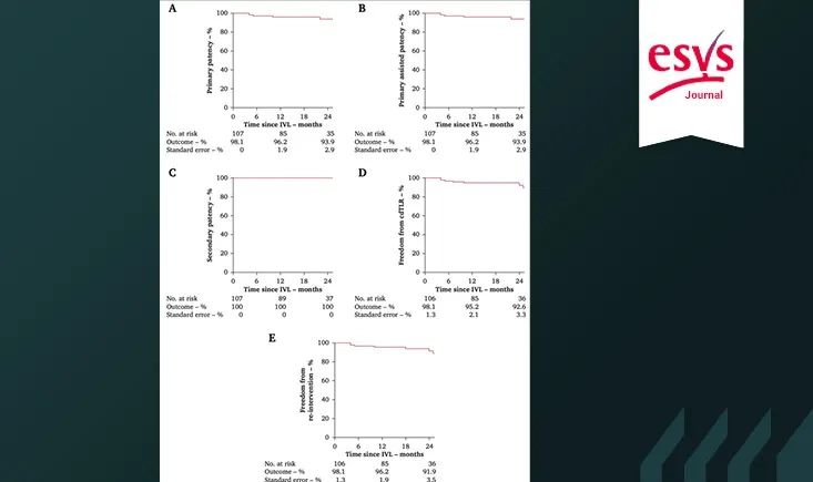
In the prospective FESTIVAL registry, IVL without stenting achieved high patency rates and low reintervention rates in patients with de novo CFA disease. Outcomes were particularly favourable in lesions not involving the femoral bifurcation.
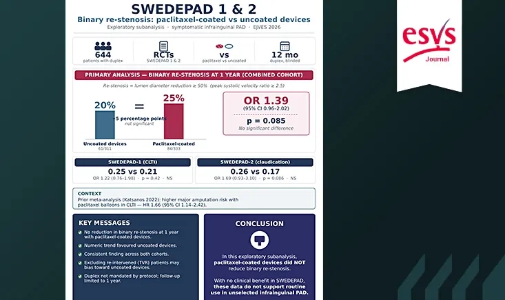
In this SWEDEPAD sub-analysis, paclitaxel-coated devices were not associated with reduced one-year binary restenosis compared with uncoated devices. These findings, consistent with the main trial results, do not support routine use in an unselected infrainguinal PAD population.
Dr Eric Steinmetz discusses key themes from the French–German vascular surgery session, including asymptomatic carotid stenosis, CREST-2 data, surgical training, and AAA repair thresholds.
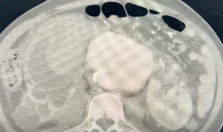
This case describes a ruptured abdominal aortic aneurysm complicated by an aortorenal fistula, presenting with massive hematuria and hemorrhagic shock in an emergency setting. It highlights an unusual clinical presentation of a life-threatening vascular pathology and the diagnostic challenges it may pose.
Dr Juliette Raffort-Lareyre interviews Dr Gilles Goyault on occlusive SFA ISR, discussing intraluminal strategies, debulking techniques, and the current lack of strong evidence.
At Charing Cross 2026, Dr Lichtenberg presented long-term data on venous stenting for acute and chronic femoral DVT. In a cohort of 80 patients followed for up to 8 years, most loss of patency occurred within the first 6 to 8 months, with early re-occlusion—likely linked to inflow issues—identified as the main cause of failure.
During a short interview recorded for PVI online at Charing Cross 2026, Prof. Secemsky discusses ongoing developments in bioresorbable scaffold technology and the key elements surrounding next-generation devices in below-the-knee intervention.
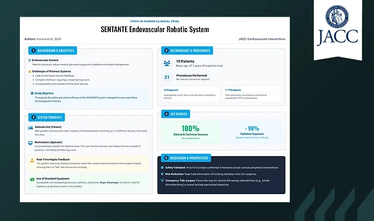
Through this publication review, discover the first-in-human evaluation of a teleoperated robotic system with haptic feedback for peripheral endovascular interventions.