H. Universitario Principe De Asturias, Madrid, SPAIN
This section provides a selection of clinical cases provided by experts in vascular techniques.
A young woman with neurofibromatosis type 1 and severe hypertension despite four antihypertensive drugs presented with a complex renal artery aneurysm. The aneurysm was relatively small, raising an important question: was its size really the main factor driving the decision to treat?


A 75-mm saccular juxtarenal aneurysm had rapidly expanded in a patient with weight loss and an inflammatory response. What treatment strategy would you choose in this challenging setting?
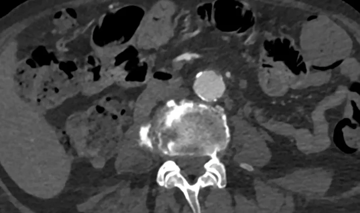

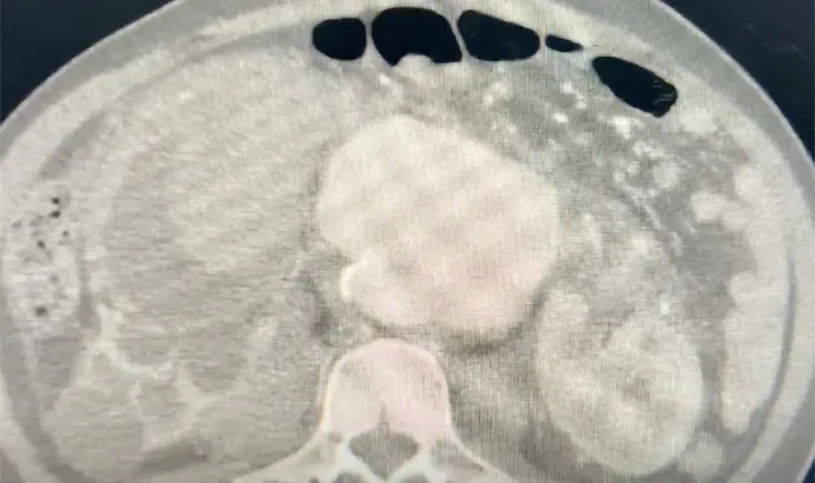
This case describes a ruptured abdominal aortic aneurysm complicated by an aortorenal fistula, presenting with massive hematuria and hemorrhagic shock in an emergency setting. It highlights an unusual clinical presentation of a life-threatening vascular pathology and the diagnostic challenges it may pose.

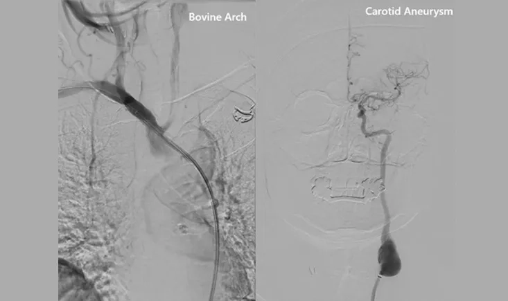
A 63-year-old man with multiple prior left carotid surgeries and a bovine aortic arch presented with a large carotid aneurysm. Open repair was high-risk, leaving the team to weigh complex anatomical and technical challenges. How should such a case be managed—and what was ultimately done?


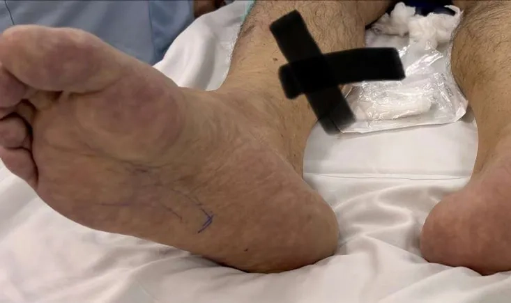
A 56-year-old patient presents with acute limb ischemia and complex multilevel arterial disease, including common femoral artery occlusion. What would be the optimal revascularisation strategy in this setting?

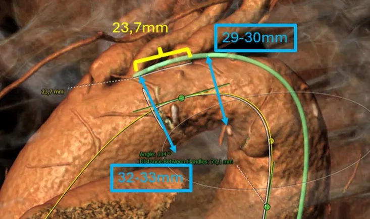
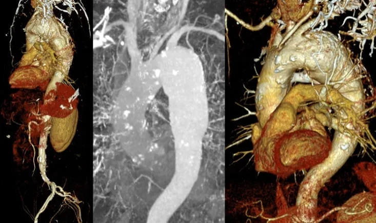
The management of aortic arch pathology is frequently demanding and poses significant anatomical challenges, ranging from open surgery to hybrid debranching procedures and, ultimately, total endovascular approaches.



This case explores the management of a unilateral total iliac artery occlusion in a high-risk patient with advanced peripheral arterial disease. Through imaging findings and clinical presentation, it invites reflection on treatment options, weighing endovascular and surgical strategies in the context of symptom severity, collateral circulation, and overall cardiovascular risk.
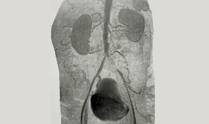
This case explores the management of a complex subclavian vein intra-stent occlusion in a haemodialysis patient, raising key questions when standard endovascular options reach their limits.
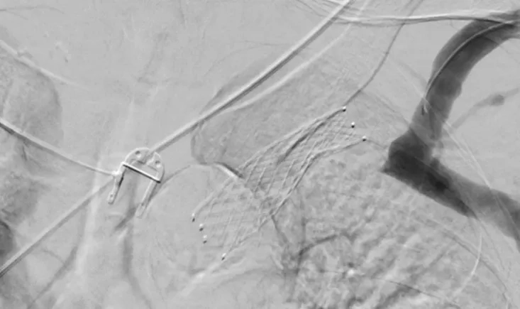




A 73-year-old man with major cardiovascular risk factors presented with acute pain and was diagnosed with a large thoraco-abdominal aneurysm and bilateral iliac aneurysms. A complex, staged endovascular repair was performed to reduce the risk of spinal cord ischemia. At 6-month follow-up, imaging revealed a type II endoleak with aneurysm sac enlargement, prompting the need for further strategic decision-making.
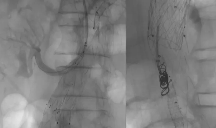



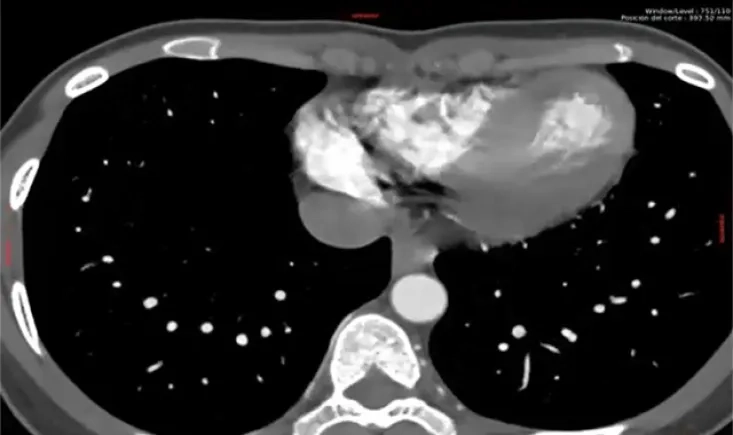
A 65-year-old male with multiple comorbidities presents with a 58 mm thoracic aortic aneurysm and a severely kinked, diseased aorta. This case reviews the key anatomical challenges, landing-zone configuration, access considerations, and protective strategies needed when planning a patient-tailored endovascular repair.